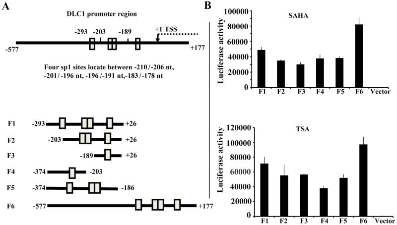
Fig. 2. SAHA and TSA activate DLC1 promoter with the same SP1 sites.
A: Schematic representation of the DLC1 promoter region, which spans from −577 to + 177 and contains four SP1sites locate between −210/−206, −201/−196, −196/−191, −183/−178. Fragments with different combinations of SP1 sites are shown in the bottom. The numbers refer to the transcription start site (left panel). B: Activation of the truncated constructs of DLC-1 promoter in 22Rv1 cells. The constructs were transiently transfected into 22Rv1 cells followed by treatment with TSA or SAHA. Luciferase activity was measured and normalized to protein concentration. Data presented are representative of three independent experiments (right panel).

