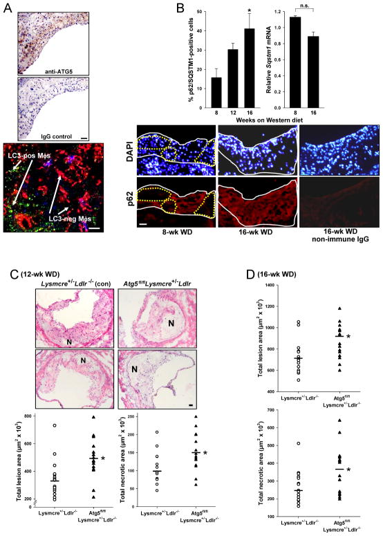
Figure 3. Defective Macrophage Autophagy Promotes Plaque Necrosis in Advanced Atherosclerotic Lesions of Western diet (WD)-Fed Ldlr−/− Mice.
(A) Female GFP-LC3-Ldlr−/− mice were fed the WD for 12 wks. Cross-sections of an aortic root lesion were immunostained with anti-ATG5 or control IgG (top images; bar = 20 μm) or with anti-F4/80 antibody and viewed by confocal fluorescence microscopy (bottom image; red = F4/80, green = GFP-LC3; bar = 10 μm).
(B) Female Lysmcre+/−Ldlr−/− (control) and Atg5fl/flLysmcre+/−Ldlr−/− mice were fed the WD for 8, 12, or 16 wks (n = 5 per group). Lesions were then immunostained for p62/SQSTM1 and DAPI (nuclei), and lesional Sqstm1 mRNA was assayed by LCM-RT-qPCR (mean ± S.E.M.; *p = 0.02 vs. 8-wk value; n.s., not significant). Representative images for the p62 immunostaining data are shown for the 8- and 16-wk lesions, along with a non-immune control image for a 16-wk lesion. Bar, 20 μm. In these images, the intima, which is composed mostly of macrophages, is outlined by the solid white lines, and areas of the 8-wk intima that have cells negative for p62 are outlined by the dotted yellow line.
(C–D) Aortic root sections of female Lysmcre+/−Ldlr−/− (control) and Atg5fl/flLysmcre+/−Ldlr−/− mice fed the WD for 12 or 16 wks were stained with H&E and then quantified for lesion and necrotic area. Representative images are shown for the 12-wk groups. N, necrotic area; Bar, 20 μm (mean ± S.E.M.; *p < 0.05; n con/experimental = 15/15 for the 12-wk cohort and 17/15 for the 16-wk cohort).

