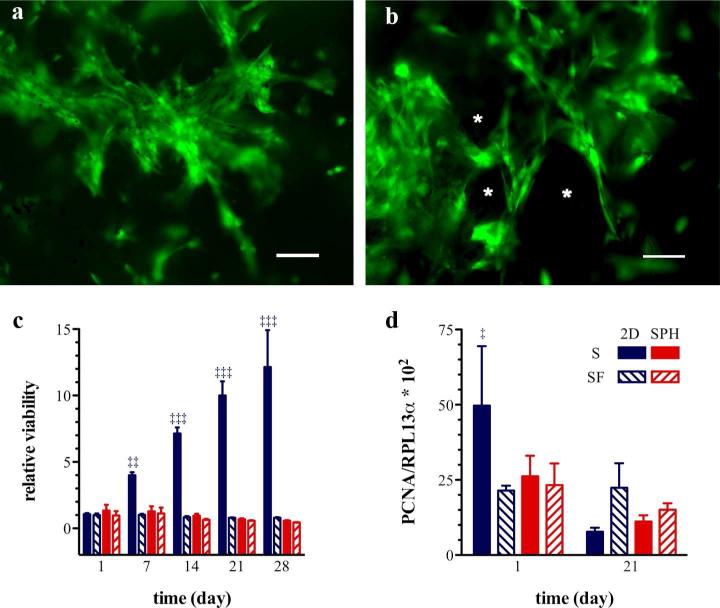
Figure 1. Human mesenchymal stem cells remain viable and proliferate within PEGDA superporous hydrogels.
Representative pseudocolored epifluorescent micrographs of viable hMSCs around the pores (*) of PEGDA SPHs, (a) 3 and (b) 7 weeks post seeding. Viable cells are stained with calcein acetoxymethyl ester dye (green) and dead cells appear red due to staining with ethidium homodimer. The scale bar is 100 μm. (c) MSC viability within PEGDA superporous hydrogels (SPH/red) and on tissue culture plastic as a monolayer (2D/blue) under serum-containing (S/solid bars) and serum-free conditions (SF/diagonal lines within bars) was assessed by the MTS assay. Cells were incubated for one day in the presence of serum, at t = 0 day, and maintained for additional time (as listed on x-axis) in either serum-containing (S) or serum-free conditions (SF). The symbols (‡‡) and (‡‡‡) denote a significant higher cell viability under 2D culture conditions in the presence of serum compared to all other groups at p < 0.01 and p < 0.001 respectively. (d) The proliferative activity of hMSCs was evaluated by the gene expression of PCNA. The symbol (‡) denotes a significant difference in the PCNA mRNA expression at day 1 under 2D culture conditions in the presence of serum compared to all other groups at p < 0.05 (n=3-4; mean ± standard deviation).

