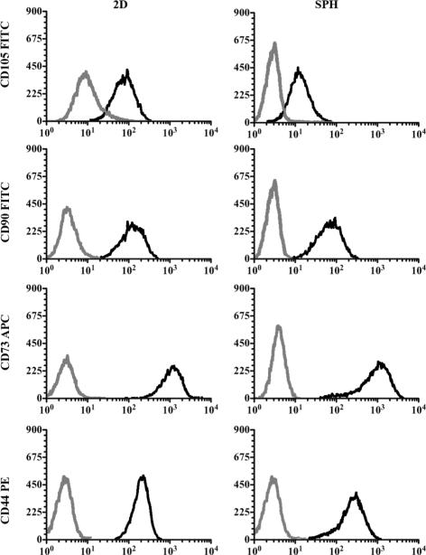
Figure 5. Expression of human mesenchymal stem cell surface markers of cells cultured within PEGDA superporous hydrogels or on tissue culture plastic (TCP) for 21 days.
Representative flow cytometry histograms from three independent experiments with three different donors are shown. Analysis revealed no statistically significant differences in the expression of CD105, CD90, CD73 and CD44 in hMSCs cultured within SPHs (right) or in 2D (left). Gray lines represent the fluorochrome- and isotype-matched control and black lines the corresponding CD marker-specific antibody.

