Figure 4.
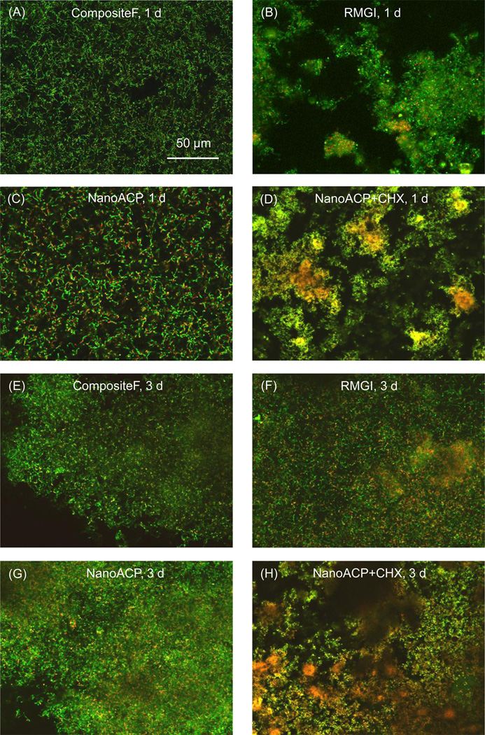
Representative live/dead images: Early biofilms (1 d) are shown for (A) CompositeF, (B) RMGI, (C) NanoACP, and (D) NanoACP+CHX. Mature biofilms (3 d) are shown for the same materials in (E) to (H). Live bacteria appear green. Membrane-compromised bacteria appear red, and, when mingled with live bacteria, appear yellow/orange. At 1 d, bacteria were predominantly alive in (A) to (C), with some cell death in (B). (D) NanoACP+CHX had increased dead bacteria relative to (A) to (C). At 3 d, live bacteria formed mature biofilms in (E) to (G) with slightly increased death in (F). (H) NanoACP+CHX had significant amounts of dead bacteria. Images of CompositeNoF and NanoCaF2, which had qualitatively similar features to the other materials without CHX, were omitted to save space. Likewise, images for NanoCaF2+CHX were not shown, as their biofilms had similar features to biofilms on the NanoACP+CHX.

