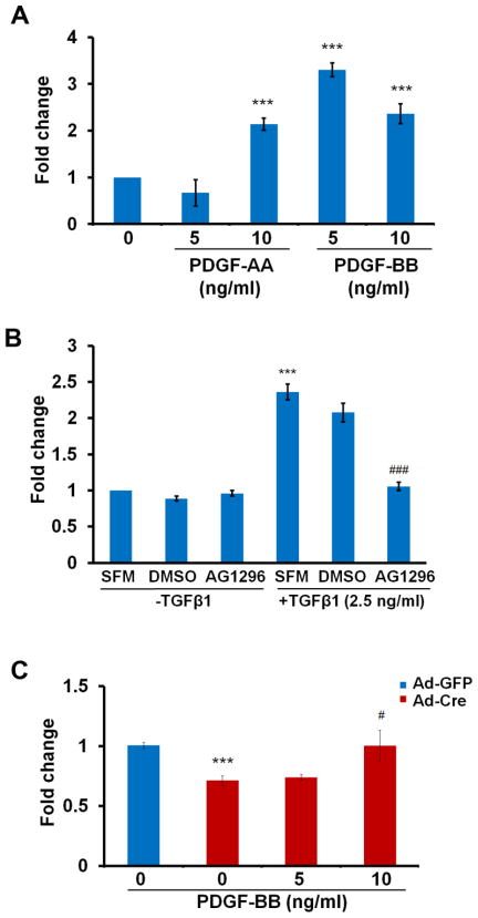
Figure 3. PDGF ligands plays an important role in TGFβ mediated cell migration.
(A) Cells were isolated from wild type embryos and were either treated or not treated with PDGF-AA or PDGF-BB at the bottom of the well. Both PDGF ligands stimulated cell migration. ***, P<0.001. (B) Cells from wild type embryos were pre-treated with AG1296 (25 μM) for 30 min. before being placed in the migration assay with or without TGFβ in the bottom well. DMSO is the solvent control. SFM is the serum free medium control. TGFβ-stimulated migration was blocked by the PDGFR antagonist. ***, P<0.001 [SFM +TGFβ1 vs SFM-TGFβ1]; ###, P<0.001 [AG1296+TGFβ1 vs DMSO +TGFβ1]. (C) Cells were isolated from Tgfbr2fl/fl embryos and infected with Ad-Cre to delete Tgfbr2 or with Ad-GFP as a control. The cells were placed in the migration assay with or without PDGF-BB in the bottom well. PDGF-BB stimulated migration in Tgfbr2 deleted cells. ***, P<0.001 [Ad-Cre vs Ad-GFP]; #, P<0.01[Ad-Cre+PDGF-BB 10ng/ml vs Ad-Cre]. Representative of two separate experiments is shown.

