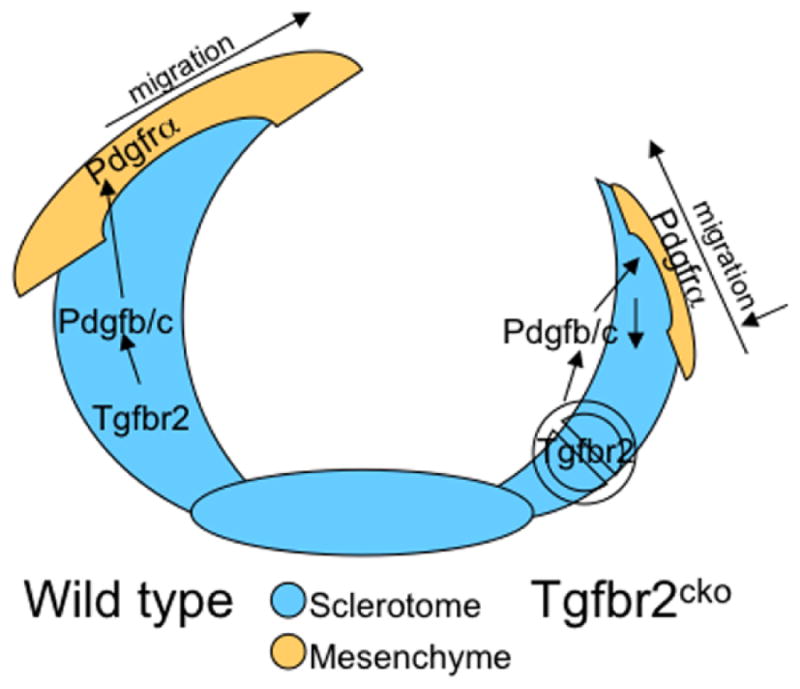
Figure 5. Proposed model for the role of Tgfbr2 in development of the spinous process.

We propose the following model for TGFβ action in the development of the spinous process. In wild type embryos (left), TGFβ acts on its receptor in cartilage derived from sclerotome to stimulate PDGF ligand expression. PDGF ligands are then secreted and bind to PDGFRα on the adjacent mesenchyme. This paracrine activation of PDGF signaling promotes proliferation and migration of these cells eventually leading to the closure of the neural arches and formation of the spinous process. In Tgfbr2cko embryos (right), PDGF ligands are not made in sufficient amount to stimulate proliferation and migration of the adjacent mesenchyme. Alterations in the adjacent mesenchyme lead to failure in the formation of the spinous process.
