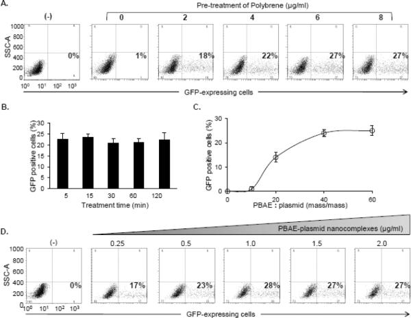
Figure 4. Optimization of the nanocomplex-mediated cell transfection.
A, Dose effect of polybrene. Jurkat cells (5×10⁶) were pre-treated with polybrene for 5 min at different concentrations (0–8 μg/mL) and then exposed to the PBAE polymer (> 7 kDa)-plasmid nanocomplex. In the control experiment, reporter plasmid DNA alone (0.25 μg/mL) was used (−). After treatment for 48 hr, GFP-expressing cells were quantified by flow cytometry (%). B, Time-course of polybrene pre-treatment. Cells were pre-treated with 6 μg/mL polybrene for different times (5–120 min) and followed by exposure to the PBAE polymer-plasmid nanocomplex. Resultant GFP-expressing cells were quantified by flow cytometry (%). C, Optimal ratio of PBAE polymers and plasmid DNA reporters in the nanocomplex. Cells were pre-treated with polybrene (6 μg/mL) for 5 min and transfected by the nanocomplexes composed of PBAE > 7 kDa and pMax-GFP at different ratios (mass/mass) as indicated. Resultant GFP-expressing cells were quantified by flow cytometry (%). D, Optimal amount of reporter gene in cell transfection. Cells were pre-treated with polybrene (6 μg/mL) for 5 min and transfected by different doses of the nanocomplexes with the fixed 40:1 ratio of PBAE polymers (> 7 kDa) and pMax-GFP. The final amounts of reporter pMax-GFP were used from 0.25 to 2.0 μg/mL. Reporter plasmid DNA alone was used in control cells (−). Resultant GFP-expressing cells were quantified by flow cytometry (%).

