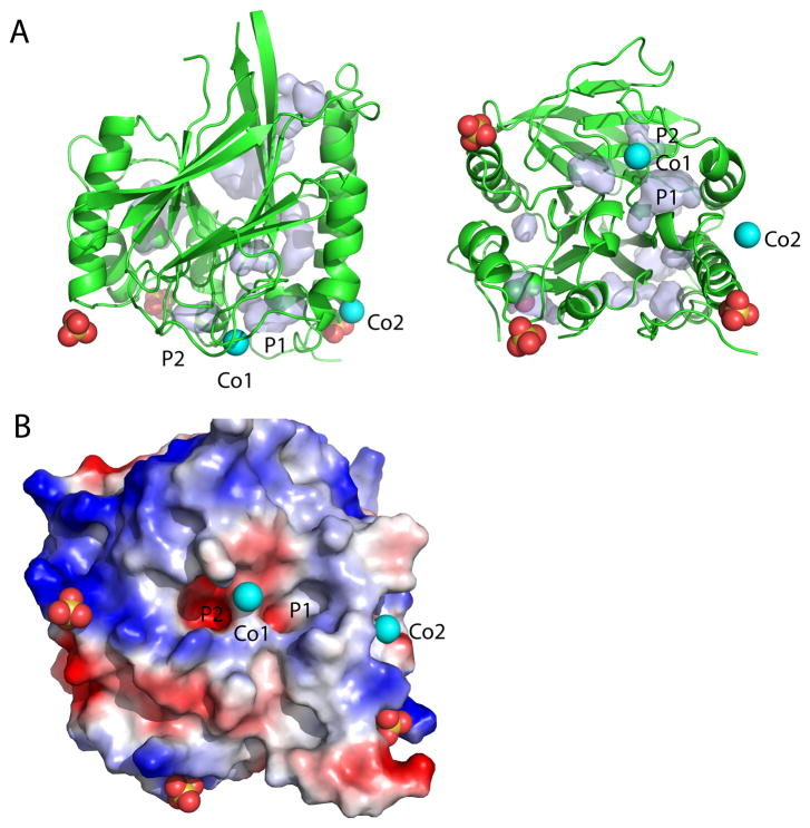
Figure 8. Pockets of GAP50 near the homologous active site region in other proteins.
A) Ribbon diagram of PfGAP50 with identified ligands shown in spheres, the orientation is identical to Figure 4A. Pockets and cavities as calculated by PyMol are as light blue surfaces. Pocket 1 (P1) and pocket 2 (P2) are close to the cobalt atom closest to the active site of the human purple acid phosphatase.
B) Electrostatic potential around pockets 1 and 2 in an orientation as viewed in Figure 4B–D, with P2 being more charged compared to P1.

