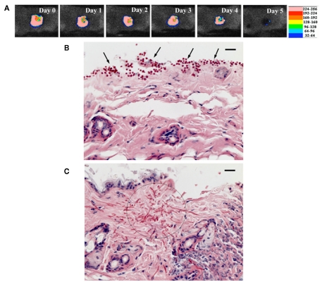
Figure 6.
(A) Successive bioluminescence images of a representative mouse skin abrasion infected with 107 CFU C. albicans. (B,C) Representative periodic acid-Schiff-stained skin biopsy specimen taken from a mouse skin abrasion infected with 107 CFU C. albicans, showing the presence of yeasts [(B), arrows], hyphal filaments [(C), circle], and inflammatory infiltrate (C). Biopsy was taken on day 1 postinfection. Bar, 20 μm.

