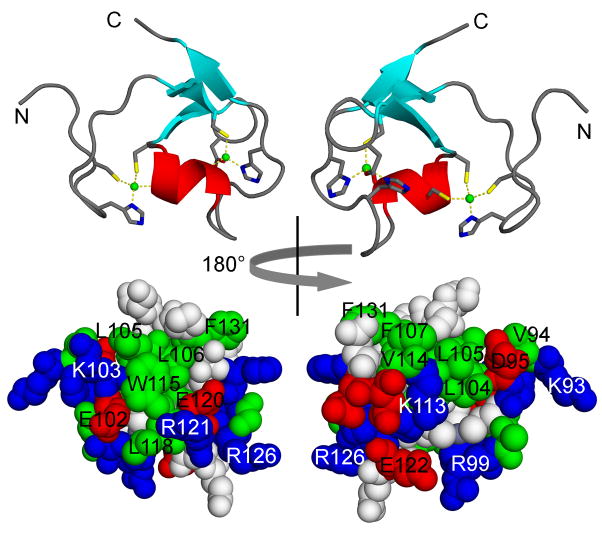
Figure 4.
NMR solution structure of human TRIM5α amino acid residues 86 to 131, encompassing the B-box 2 domain [58].
Top: ribbon diagrams with Zn2+ ions indicated as green balls, in a CHCDC2H2 coordination mode. Coordinating residues are highlighted.
Bottom: surface representations.
Left: hydrophobic surface cluster 1.
Right: hydrophobic surface cluster 2.

