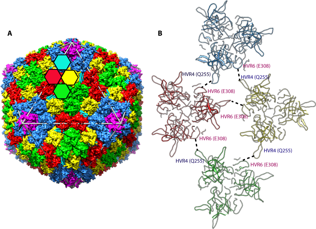
Figure 1.

A. A schematic representation illustrating the arrangement of major capsid proteins (hexon and penton base) in the adenovirus capsid. The colored hexagons represent the 4 unique hexon trimers of the pseudo T=25 icosahedral lattice. Five-fold related penton base subunits shown in magenta are located at each of the 12 vertices of the capsid. B. Interaction of hypervariable (HVR) loops HVR4 and HVR6 that help stabilize the Ad capsid. These interactions result in the ordering of HVR4 loop, which was disordered in the isolated hexon structure.
