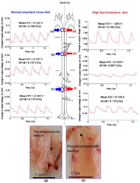
FIGURE 4.
Intravascular voltage (IVV) signals in the rabbit aortas on normal standard (ND) vs. high-fat (HD) diets. (a–c) Representative intravascular voltage (IVV) profiles were acquired in the distal aortic arch, thoracic aorta, and abdominal aorta on ND. (d–f) Representative IVV profiles were acquired in the HD group. (g, h) Gross pathology revealed pre-atherosclerotic lesions in both distal aortic arch and thoracic aortas from the HD group.

