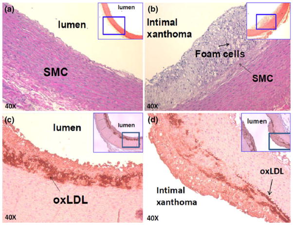
FIGURE 5.
Immunostaining of pre-atherosclerotic lesions on ND vs. HD. (a) Aortic arch isolated from the rabbit on ND. (b) Intimal thickening and atheroma were present in the fat-fed rabbits. Macrophage-derived foam cells were identified Sudan black stain, and varying degrees of smooth muscle cells (SMC) were identified by anti-SMC actin. (c, d) Subendothelial layers were stained positive for oxLDL by mAb4E6 antibody.

