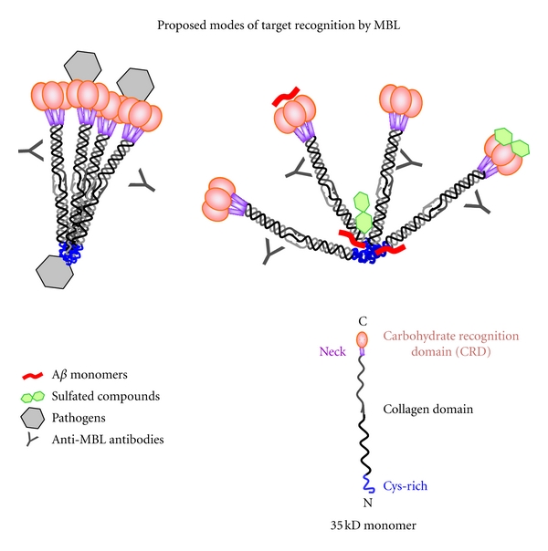
Figure 7.

A proposed model of MBL-ligand-binding modes. Figures on the right and left both represent tetramers of MBL trimers. On the left, the MBL molecule is shown binding to pathogens by both the CRD and CysD regions. On the right, sulfated carbohydrates are depicted binding to both the CRD and CysD regions, which may inhibit binding of ligands, including Aβ, to the CysD. Additionally, the CRDs may have alternative bindings modes, which allow recognition of carbohydrate and noncarbohydrates, possibly including Aβ. Antibodies are shown binding to the collagen domain of MBL, corresponding to the anti-MBL antibodies used in the ELISA experiments reported here.
