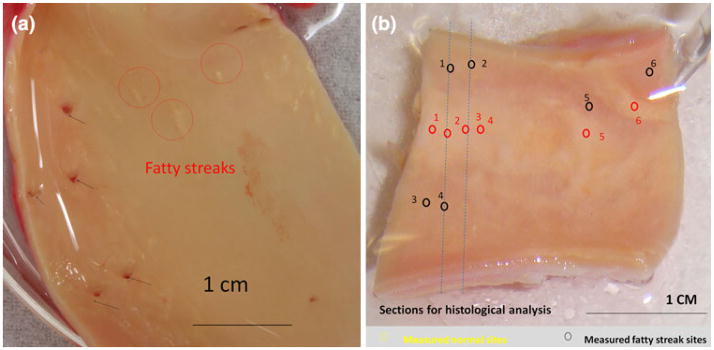
FIGURE 3.
Ex vivo segments of human descending thoracic aorta and common carotid artery. (a) En face thoracic aorta, revealing several orifices to the intercostal branches (black arrows) and fatty streaks (red circles) corresponding to the EIS readouts in Fig. 7a. (b) Numerous fatty streaks (red circles) and apparently normal endothelium (black circles) corresponded to the EIS readouts in Fig. 8a. Cross-sectional histological analyses were performed along blue dashed lines (Figs. 8c and 8d).

