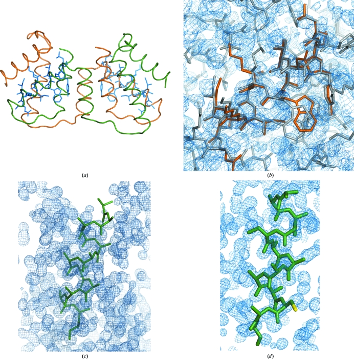
Figure 3.
Structure solution with fragments with side chains. (a) Final structure of PRD2 shown as a backbone trace, with superimposed helices with side chains modelled in standard conformations, as located in the successful solution. (b) Detailed view of one of these helices superimposed on the final structure and the resulting electron-density map after SHELXE expansion contoured at 1σ and including data extrapolated to 1.7 Å. (c) Slightly misplaced polyalanine helix that nevertheless leads to structure solution in the case of viscotoxin A1. (d) Helix with cysteine side chain (yellow) in standard conformation used in the case of viscotoxin A1. In this case part of the helix is also misplaced, including the cysteine side chain, but this substructure also leads to a final electron-density map shown in cyan contoured at 1σ, including data extrapolated to 1.0 Å and characterized by a mean phase error of 18.9°. Figures were prepared with PyMOL and Coot (DeLano, 2002 ▶; Emsley et al., 2010 ▶).

