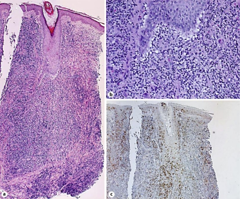
Fig. 2.
A dense lymphocytic infiltrate containing numerous histiocytes that surrounded and infiltrated hypertrophic hair follicles. The infiltrate is separated from the epidermis by a grenz zone. There was no reactive pattern to the follicular centers. (a). Infiltrated cells were medium-sized with a high nuclear/cytoplasmic ratio and prominent nucleoli (b). CD1a-positive cells were densely infiltrated around the hair follicle (c). Original magnification ×50 (a, c), ×200 (b).

