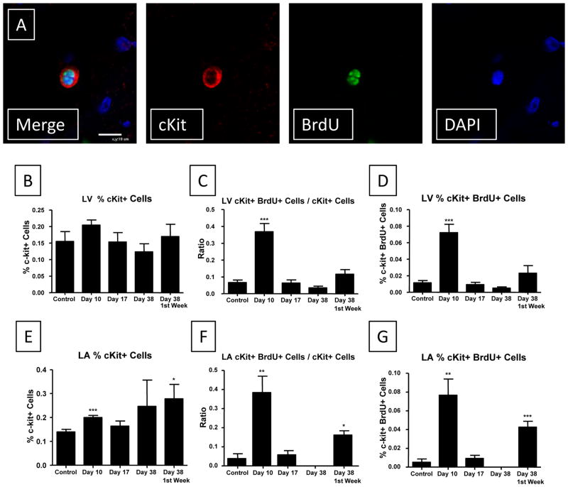
Figure 7. cKit+ cells in the heart proliferate during ISO induced injury.
A: Representative cKit staining of a proliferative cell is shown. B: The % of cKit cells in the ventricle did not change during the time course of our study. C: A significant increase in the ratio of proliferative cKit cells to non-proliferative cKit cells was observed during the injury phase. D: cKit+/BrdU+ cells are a very small % of the cells in the ventricle, but this % increases during ISO injury. E–G: Similar results were observed in the left atrial sections. *P<0.05; **P<0.01; ***P<0.001 versus Control. Data are presented as mean ± SEM.

