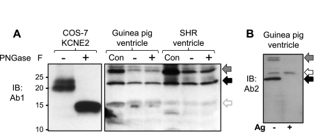
Fig. 2.
Ab1 and Ab2 detect similar banding patterns of native KCNE2 proteins in SHR and guinea pig hearts, which are not N-glycosylated. A: PNGase F treatment collapses the ≥20-kDa KCNE2 bands expressed in COS-7 cells into the core unglycosylated 15-kDa band, but does not alter the banding pattern of native proteins in guinea pig and SHR ventricles detected by Ab1. Con, original WTL; +, PNGase F treated; −, similarly processed in the absence of enzyme. B: WTL from guinea pig ventricle probed with Ab2, without or with preincubation of Ab2 with excess Ag (− and +, marked below the image). In both A and B, the solid back arrows point to the 24-kDa native KCNE2 band and arrows with gradient gray point to the 32-kDa putative KCNE2 band. In A, the open gray arrow points to the 15-kDa unglycosylated KCNE2 band. In B, the open black arrow points to a band detected by Ab2 that likely represents an unrelated protein.

