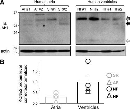
Fig. 5.
Quantifying KCNE2 in 4 human atrial specimens [2 from patients in atrial fibrillation and 2 from patients in sinus rhythm (AF and SR), respectively] and 4 human ventricular specimens [2 from nonfailing and 2 from failing hearts (NF and HF), respectively]. A: membrane is probed for KCNE2 (Ab1), stripped, and reprobed for actin. Coomassie blue (CB) stain confirms even loading (not shown). B: densitometry quantification of KCNE2 protein level. Data analysis is the same as that described for Fig. 3B. Mean values are shown as histogram bars with SE, and data from individual specimens are shown as symbols.

