Figure.
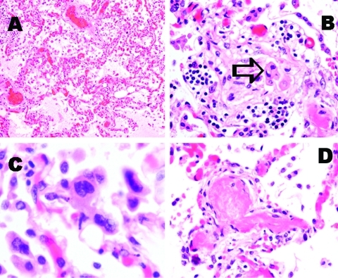
Pathologic findings of lung tissue sections. A: Pulmonary congestion and edema (H&E stain, original magnification x100). B: A mild degree of interstitial lymphocytic infiltration. Intra-alveolar organizing exudative lesion was occasionally found. Detached atypical pneumocytes indicated by arrow (H&E stain, original magnification x200). C: Atypical multinucleated pneumocytes were occasionally identified. Definite viral inclusion was not apparent (H&E stain, original magnification x400). D: Fibrin thrombi were frequently noted in small pulmonary arteries and arterioles (H&E stain, original magnification x200).

