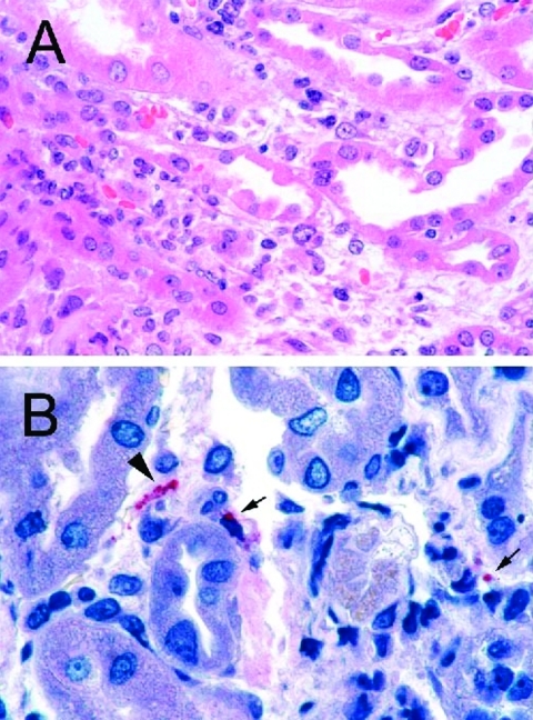
Figure 2.
A: Renal biopsy shows inflammatory cell infiltrate in the interstitium and focal denudation of tubular epithelial cells. Hematoxylin and Eosin; original magnifications x100. B: Immunostaining of fragmented leptospire (arrowhead) and granular form of bacterial antigens (arrows). Original magnifications x158.

