FIGURE 4.
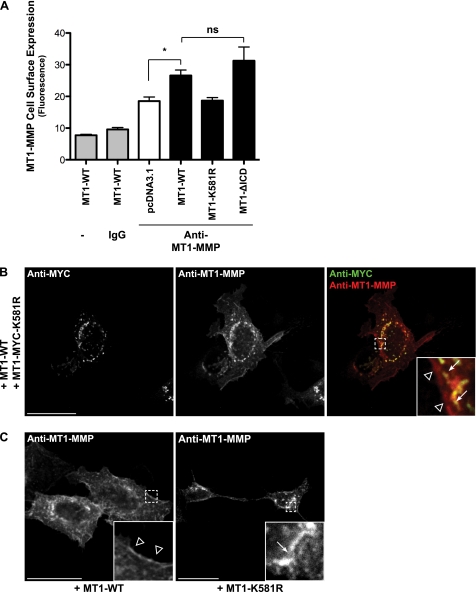
Mutation of the MT1-MMP ubiquitination site at Lys581 decreases MT1-MMP cell surface expression. A, MCF-7 cells expressing pcDNA3.1, MT1-WT, MT1-K581R, or MT1-ΔICD were fixed, and MT1-MMP cell surface expression was detected by flow cytometry with an antibody against the MT1-MMP catalytic domain (LEM-2/15.8). Furthermore, MT1-WT expressing cells remained unstained as control. Data represent the mean fluorescence (n = 3 ± S.E.) with p < 0.05 (*). ns, not significant. B and C, MCF-7 cells were either co-transfected with MT1-WT and MT1-MYC-K581R (B) or MT1-WT and MT1-K581R were transfected individually (C). Cells were subsequently fixed, permeabilized, and immunostained with an anti-Myc (Alexa Fluor® 488 secondary, green) and an anti-MT1-MMP antibody (N175/6; Cy5 secondary, red) (B) or with an anti-MT1-MMP antibody (N175/6; Alexa Fluor 488® secondary, green) (C). Arrows point at intracellular staining of the MT1-K581R mutants, whereas arrowheads indicate staining at or close to the plasma membrane of MT1-WT. Bars represent a distance of 25 μm.

