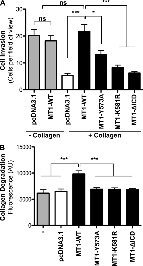
FIGURE 6.
Ablation of MT1-MMP ubiquitination reduces cell invasion through type I collagen. A, MCF-7 cells were transfected with either pcDNA3.1, MT1-WT, MT1-Y573A, MT1-K581R, or MT1-ΔICD, and cell migration was quantified using a modified Boyden chamber assay. Data represent mean number of cells per field of view of three independent biological replicates with p < 0.0001 (***) and p < 0.01 (*). ns, not significant. B, wild-type MCF-7 cells or MCF-7 cells expressing pcDNA3.1, MT1-WT, MT1-Y573A, MT1-K581R, or MT1-ΔICD were co-polymerized with 1.5 mg/ml non-labeled rat tail type I collagen mixed with 2% FITC-conjugated bovine type I collagen. Following a 40-h incubation, the fluorescence of the supernatant was determined. Data represent mean fluorescence of five independent biological replicates with p < 0.0001 (***). AU, arbitrary units.

