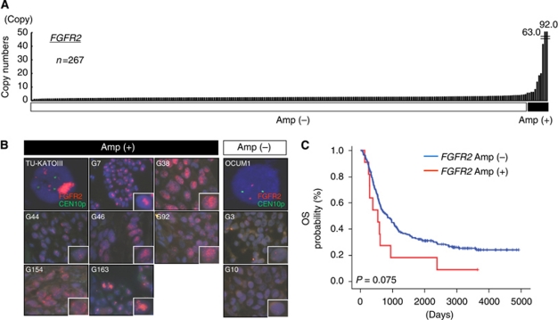
Figure 2.
(A) Amplification of FGFRs in surgical specimens of gastric cancer. A TaqMan copy number assay for FGFR2 was performed using DNA samples obtained from 267 FFPE samples. Human normal genomic DNA was used as a normal control. FGFR2 amplification over 5 copies was observed in 11 cases (92.0, 63.0, 41.4, 19.9, 18.4, 13.7, 8.3, 6.2, 6.2, 5.7 and 5.6 copies). (B) Fluorescence in situ hybridisation analysis of FGFR2-amplified KATO-III cells, non-amplified OCUM1 cells and nine surgical specimens of gastric cancer. Green, signal of CEN10P locus; Red, signal of FGFR2 locus; G3∼G92, sample numbers; Amp, gene amplification. High-power images are presented for a single cancer cell. (C) Overall survival in FGFR2-amplified gastric cancer. Kaplan–Meier curves for OS according to the FGFR2 amplification status.

