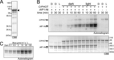
FIGURE 4.
In vitro phosphorylation assay of the full-length CrPHOT. A, 10.0% SDS-polyacrylamide gel pattern of full-length CrPHOT stained with CBB. The sample with an N-terminal His6 tag was successively purified by Ni2+-ion affinity chromatography and size-exclusion chromatography. The triangle indicates the position of the intact CrPHOT. B, in vitro phosphorylation assay for CrPHOT under BL (100 μmol m−2 s−1) or in the dark. CrPHOT and/or AtP1-Nt was incubated with radioactive ATP for the indicated durations. The upper and lower panels show the 10.0% SDS-PAGE gel patterns visualized by autoradiography and CBB staining, respectively. + or −, the presence or absence of the kinase and/or the substrate, respectively; D, dark condition; L, BL irradiation. C, dependence of the kinase activation on the light intensity. In vitro phosphorylation assay of CrPHOT was performed as described for B. The samples were incubated for 10 min under BL at the indicated light intensities. Phosphorylated AtP1-Nt was visualized by autoradiography.

