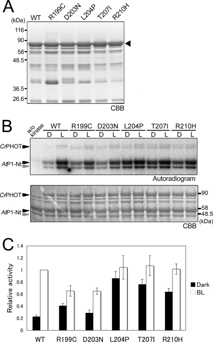
FIGURE 5.
Effects of CrPHOT mutations on in vitro phosphorylation activity. A, 10.0% SDS-polyacrylamide gel patterns of the mutant CrPHOT proteins stained with CBB. The wild-type (WT) and the mutant CrPHOT proteins were prepared as for Fig. 4A. The triangle indicates the position of the intact CrPHOT. B, in vitro phosphorylation assay for the WT and mutant CrPHOT samples. The reaction proceeded for 30 min under BL (100 μmol m−2 s−1) or in the dark condition. Sample preparation and experimental procedures were the same as those for Fig. 4B. The upper and lower panels show the 10.0% SDS-polyacrylamide gel patterns visualized by autoradiography and CBB staining, respectively. C, quantification of the phosphorylation activity in the WT and the mutant CrPHOT. An in vitro phosphorylation assay was performed as for B. Band intensities were quantified by a bioimaging analyzer and expressed relative to the maximal phosphorylation level in the WT under BL. The data represent the means of three independent experiments. Error bars, S.D.

