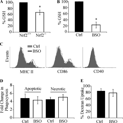
FIGURE 2.
Dysregulation in function of Nrf2−/− DC is not a consequence of lower GSH levels. A and B, GSH levels were measured in Nrf2+/+ and Nrf2−/− iDC (A) or Nrf2+/+ iDCs untreated (Control) or treated with BSO (100 μm) (B) for 24 h. Data are presented as average percentage (when compared with Nrf2+/+ or untreated iDCs) ± S.D. Statistical significance was tested by unpaired Student's t test (*, p < 0.05). Data are derived from five independent experiments. C, immature Nrf2+/+ DCs untreated or treated with BSO were labeled with antibodies against MHC II, CD86, and CD40 co-stimulatory receptors. Co-stimulatory receptor expression was determined by flow cytometry. Histogram overlays show fluorescence intensity of respective co-stimulatory receptors and are representative of four independent experiments. D, immature Nrf2+/+ DCs untreated or treated with BSO were co-cultured with CFSE-labeled apoptotic thymocytes or necrotic Jurkat cells at 37 °C for 2 h. DC phagocytic capacity was measured by flow cytometry as an increase in CFSE levels when compared with corresponding 4 °C baseline control samples. Data are presented as average -fold changes ± S.E. Data are derived from five independent experiments. E, endocytic capacity was measured by incubating Nrf2+/+ iDCs that were untreated or treated with BSO with DextranFITC for 60 min at 37 °C. DextranFITC uptake by DCs was assessed by flow cytometry. Data are derived from four independent experiments and presented as average percentage of uptake ± S.D.

