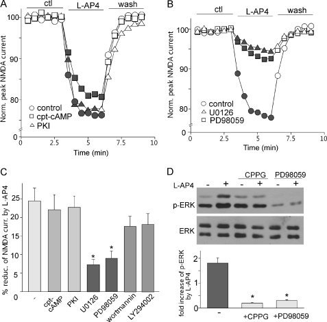
FIGURE 3.
Activation of ERK is required for mGluR7 reduction of NMDAR currents. A, plots of normalized (Norm.) peak NMDAR currents in dissociated PFC pyramidal neurons showing the effect of l-AP4 (200 μm) in the absence or presence of PKA activator cpt-cAMP (100 μm) or PKA inhibitor PKI14–22 (PKI, 0.1 μm, myristoylated). ctl, control. B, plots of normalized peak NMDAR currents showing the effect of l-AP4 (200 μm) in the absence or presence of ERK kinase inhibitors U0126 (20 μm) or PD98059 (50 μm). C, cumulative data (mean ± S.E.) showing the percentage of reduction (% reduc.) of NMDAR currents (curr.) by l-AP4 in the presence of cpt-cAMP, PKI14–22, U0126, PD98059, wortmannin (1 μm), or LY294002 (100 μm). *, p < 0.001, ANOVA. D, top, Western blotting of phospho-ERK (p-ERK) and total ERK in cultured PFC neurons showing the effect of l-AP4 in the absence or presence of CPPG or PD98059. Bottom, quantitative analysis showing the -fold increase of phospho-ERK by l-AP4 under different conditions. *, p < 0.001, ANOVA.

