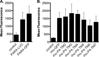
FIGURE 3.
Rhodopsin-PAR4 chimeras were expressed on the cell surface. HEK293 cells were transfected with 0.5 μg of plasmid, and cells were prepared for flow cytometry. A, PAR4 was detected with a polyclonal antibody (5 μg/ml) and anti-goat secondary antibody conjugated to Alexa Fluor647 (2 μg/ml). B, rhodopsin and rhodopsin-PAR4 chimeras were detected with a monoclonal antibody to rhodopsin (B6–30N) 1:10 dilution and anti-mouse secondary antibody conjugated to Alexa Fluor 647 (2 μg/ml). Control experiments used mock transfected cells followed by staining with anti-PAR4 or anti-rhodopsin. The data are from three or four independent experiments, and error bars represent the standard deviation.

