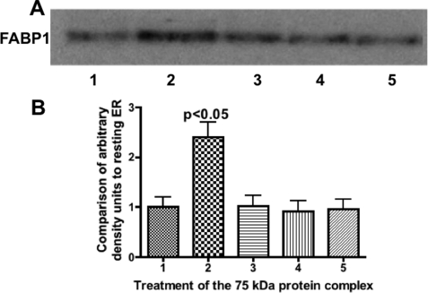
Fig. 9.
FABP1, as a member of the 75-kDa complex, has increased binding to intestinal ER only when the complex is incubated with ATP and PKCζ. Intestinal ER (300 μg) was incubated with the 75-kDa protein complex isolated from the anti-FABP1 adsorption column (50 μg). Before incubation with the ER, the complex was treated under various conditions as indicated. After the ER complex incubation, 30 μg of protein was separated by SDS-PAGE and immunoblotted using anti-FABP1 antibodies. Detection was by ECL. Lane 1, native ER. Lane 2, protein complex incubated with ATP and PKCζ at 37 °C. Lane 3, complex incubated with ATP and PKCζ at 4 °C. Lane 4, complex incubated with ATP but without PKCζ at 37 °C. Lane 5, complex incubated with PKCζ but without ATP at 37 °C. A, anti-FABP1 immunoblot. The FABP1 band is indicated. B, band densities from A were analyzed by the Gel Doc XR and recorded as densities compared with the band density of resting ER. The bars are derived from the immunoblot densities in A. The results are the mean ± S.E. of four trials The p value of test differences between the means of lanes 1, 3, 4, and 5 with lane 2 is shown.

