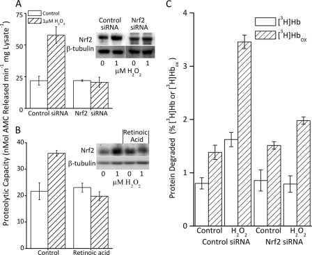
FIGURE 3.
Increased proteolytic capacity is blocked by inhibition of Nrf2. A, the increase in proteolytic capacity caused by H2O2 treatment was blocked by inhibition of Nrf2 expression through pretreatment with Nrf2 siRNA. MEF cells were grown to 10% confluence and treated with either control or Nrf2 siRNA for 24 h as described under “Experimental Procedures.” Then, 4 h after initiation of siRNA treatment, half the cells were exposed to 1 μm H2O2 for 1 h, washed, and resuspended (see ”Experimental Procedures“). The capacity to degrade the fluorogenic peptide Suc-LLVY-AMC was determined 24 h after initiation of siRNA treatment (see ”Experimental Procedures“). Values are mean ± S.E., n = 3. The inset to panel A shows a representative Western blot. B, treatment of cells with the Nrf2 inhibitor retinoic acid also blocked the H2O2-induced increase in proteolytic capacity. MEF cells were seeded at 5% confluence and treated with 3 μm retinoic acid. When cells reached 10% confluence one-half were exposed to 1 μm H2O2 and proteolytic capacity (Suc-LLVY-AMC lysis) was determined 24 h after treatment as in panel A. Values are mean ± S.E., n = 3. The inset to panel B shows a representative Western blot. C, the H2O2-induced increase in selective capacity to degrade oxidized proteins is also blocked by inhibition of Nrf2 expression. MEF cells were prepared and lysed as described in panels A and B. Lysates were incubated for 4 h with [3H]Hb or [3H]Hbox. Percent protein degraded was calculated after addition of 20% TCA and 3% BSA, and centrifugation to precipitate the remaining intact proteins (5, 12, 15, 26). Percent protein degradation was determined by release of acid soluble counts in TCA supernatants, by liquid scintillation as follows: % degradation = (acid soluble counts − background counts)/total counts × 100. Results are mean ± S.E., n = 3.

