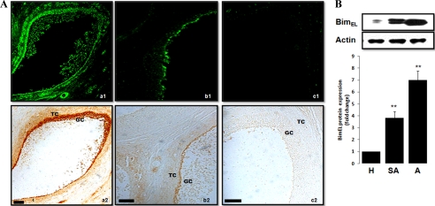
FIGURE 2.
BimEL expression pattern in granulosa cells from healthy and atretic porcine follicles. A, photomicrographs of representative examples of TUNEL and BimEL immunostaining in the porcine atretic (panel a), slightly atretic (panel b), and healthy (panel c) follicles. Cell death was detected by TUNEL (panels a1, b1, and c1), and on a consecutive section, BimEL proteins (panels a2, b2, and c2) were localized with specific antibodies by the diaminobenzidine (DAB) method. GC, granulosa cells; TC, theca cells. Bar, 50 μm. B, isolated granulosa cell lysates, obtained from follicle of different health status (H, healthy follicle; SA, slightly atretic follicle; A, atretic follicle), were subjected to SDS-PAGE and immunoblotting using anti-Bim antibodies. Actin was used as fractionation control. A representative blot is shown. The graph demonstrates the results of the densitometric analysis of BimEL protein levels normalized against loading controls (arbitrary units, healthy follicle = 1). Values represent means ± S.E. of three experiments. **, p < 0.01.

