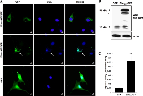
FIGURE 3.
Overexpression of porcine BimEL induces apoptosis of granulosa cells in vitro. A, cells plated on glass bottom 35-mm dishes were 80% confluent when transfected with pEGFP-N1-BimEL (panels a and b) or pEGFP-N1 (panel c, control) constructs. Note that BimEL-GFP was localized to the cytoplasm at 2 h post-transfection, and apoptotic phenotypes measured using Hoechst 33342 staining could be found in pEGFP-N1-BimEL-transfected cells at 8 h post-transfection (arrow). However, GFP-only expressing cells consistently had intact nuclei, and GFP was distributed throughout the cells. B, Western blot (WB) analysis of protein expression for endogenous and exogenous BimEL in granulosa cells transfected with a full-length BimEL-GFP fusion construct or mock vector. Transfected granulosa cells lysates were subjected to SDS-PAGE and immunoblotting using the anti-Bim antibody. Molecular masses of the protein bands are shown on the left side. Actin was used as fractionation control. * indicates an alternative splicing form of BimEL. C, caspase-3 activity was determined by caspase assay in GFP only or BimEL-GFP transfected granulosa cells (n = 4). Values represent means ± S.E. of three experiments. **, p < 0.01.

