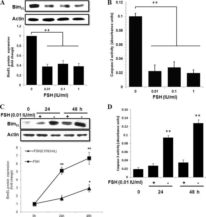
FIGURE 4.
Effect of FSH on apoptosis and BimEL expression in cultured primary granulosa cells. A, granulosa cells were obtained from porcine healthy and 2–5 mm in diameter follicles and cultured for 24 h without or with 0.01, 0.1, and 1 IU/ml porcine FSH. C, granulosa cells cultured without or with 0.01 IU/ml porcine FSH for the indicated times. BimEL protein levels were assessed by immunoblotting. Actin was used as fractionation control. A representative blot is shown. The graph demonstrates the results of the densitometric analysis of BimEL protein levels normalized against loading controls (arbitrary units, without FSH and FSH 0 h = 1, respectively). Values represent means ± S.E. of three experiments. * p < 0.05, **, p < 0.01. B and D, in parallel experiments, granulosa cells from different treatment were harvested to caspase-3 activity assays. **, p < 0.01.

