FIGURE 5.
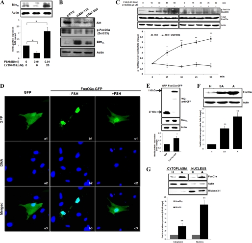
PI3K/Akt/FoxO3a pathway is involved in FSH regulation of BimEL expression. A, granulosa cells preincubated with or without 20 μm LY294002 for 30 min were treated with FSH for 16 h. The LY294002 remained present throughout the FSH stimulation. BimEL expression was assessed by immunoblotting. The graph demonstrates the results of the densitometric analysis of BimEL protein levels normalized against loading controls (arbitrary units, no FSH and LY294002 = 1). B, effect of Akt siRNA on BimEL expression. Granulosa cells were transfected with Akt siRNA (siAkt) or siCTR for 36 h and then were harvested and analyzed by immunoblotting. C, Western blot analysis was completed to determine whether FSH affected FoxO3a phosphorylation status. Granulosa cells were preincubated for 30 min with 20 μm LY294002 or not and then were harvested at specific times after FSH treatment (15–90 min). Phosphorylated (p) Foxo3a and total Foxo3a expression was assessed by immunoblotting. The graph demonstrates the ratio of the densitometric analysis of phosphorylated forms of FoxO3a proteins normalized against its total forms (arbitrary units, 0 h = 1, respectively). D, FSH regulates FoxO3a protein subcellular localization. Granulosa cells plated on glass bottom 35-mm dishes were 80% confluent when transfected with pEGFP-N1 (panel a, control) or pEGFP-N1-FoxO3a (panels b and c) constructs incubated with or without FSH. At 6 h post-transfection, cells were then stained with Hoechst 33342 to visualize nuclei, and the subcellular localization of FoxO3a was monitored by GFP fluorescence. E, Western blot (WB) analysis of fusion protein expression in granulosa cells transfected with a full-length BimEL-GFP construct or mock vector. Transfected granulosa cells lysates were subjected to SDS-PAGE and immunoblotting using the anti-GFP antibody. Molecular masses of the protein bands are shown on the left side. The graph demonstrates the results of the densitometric analysis of BimEL protein levels normalized against loading controls (arbitrary units, GFP = 1). F, FoxO3a is up-regulated in granulosa cells from atretic follicles. Isolated granulosa cell lysates, obtained from follicle of different health status (H, healthy follicle; SA, slightly atretic follicle; A, atretic follicle), were subjected to SDS-PAGE and immunoblotting using anti-FoxO3a antibodies. The graph demonstrates the results of the densitometric analysis of FoxO3a protein levels normalized against loading controls (arbitrary units, H = 1). G, immunoblots showing the expression of FoxO3a in cytosolic and nuclear extracts of granulosa cells from the healthy and atretic follicles. Actin and histone 3.1 were used as fractionation controls, respectively. The graph demonstrates the results of the densitometric analysis of FoxO3a protein levels normalized against loading controls (arbitrary units, H = 1). All values represent means ± S.E. of three experiments. *, p < 0.05; **, p < 0.01.

