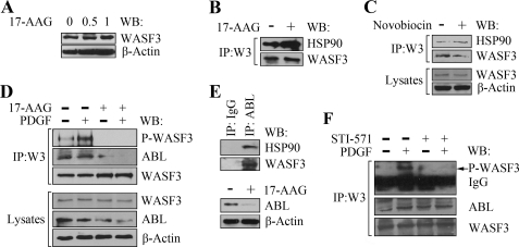
FIGURE 2.
Inhibition of HSP90 leads to WASF3 inactivation. A, treatment of PC3 cells with varying concentrations (μm) of 17-AAG for 24 h does not affect intracellular WASF3 protein levels. B, IP of WASF3 from PC3 cells treated with 17-AAG for 24 h demonstrates increased HSP90 levels in the immunocomplex compared with untreated cells. C, treatment of PC3 cells with 100 nm novobiocin for 24 h shows a decrease in WASF3 levels and a slight increase in HSP90 levels in the immunocomplex recovered following IP with an anti-WASF3 antibody. D, Western blot (WB) analysis of WASF3 IPs (upper panel) shows increased levels of phosphoactivated WASF3 in serum-starved PC3 cells using anti-phosphotyrosine antibodies (PY20) following stimulation with 50 ng/ml PDGF for 10 min. After a 24-h exposure to 1 μm 17-AAG, phosphoactivated WASF3 is undetectable despite high levels of WASF3 protein. This loss of activated protein cannot be recovered following PDGF treatment. IP of WASF3 also results in decreased levels of ABL in the immunocomplex after 17-AAG treatment regardless of PDGF stimulation. E, Western blot analysis of the ABL immunocomplex shows the presence of HSP90 in PC3 cells. Following treatment with 17-AAG levels of ABL are reduced. F, IP of WASF3 from PC3 cells shows increased activation following treatment with PDGF. In the presence of 50 μm ABL inhibitor STI571, this activation is significantly reduced and cannot be recovered in the presence of PDGF.

