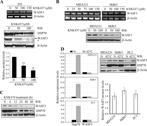
FIGURE 4.
HSP70 stabilizes the WASF3 protein. Treatment of HEK293 cells with varying concentrations of KNK437 for 24 h (A) demonstrates no effect on WASF3 mRNA levels by RT-PCR (upper panel). WASF3 protein levels determined by Western blot analysis (lower panel), demonstrates significantly reduced WASF3 protein levels (middle panel). Quantification of the Western blot data for WASF3 using ImageJ software is shown in the lower panel. **, p = 0.001. The same effects of KNK437 treatment was seen for MDA-MB-231 and SkBr3 cells (B) using RT-PCR (upper panel) and Western blot (WB) analysis (lower panel). C, when MDA-MB-231 cells were treated with 100 μm KNK437 for varying times (0–60 h), Western blot analysis of the WASF3 protein using an anti-WASF3 antibody, shows an increasing reduction in both WASF3 and HSP70 proteins. D, quantitative RT-PCR analysis (left panel) of MDA-MB-231, SkBr3, and PC3 cells exposed to heat shock (42 °C for 1 h) show large increases in HSP70 expression levels but not WASF3 (N, 37 °C treatment). Western blot analysis of the same extracts (right panel) shows that the increased HSP70 protein levels are accompanied by increased levels of WASF3. Quantification of the 37 °C/42 °C ratios using ImageJ software for WASF3 protein levels from two independent experiments are shown on the lower right panel.

