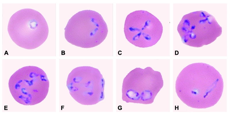
Figure 2.
Panel of Babesia-infected erythrocytes photographed from pretreatment, Wrights-Giemsa–stained smears of fresh blood obtained from the patient on July 31, 2002. The mean corpuscular volume of the erythrocytes was 103 (normal range 80–100 μm3). Note the multiply infected erythrocytes; the pleomorphism of the parasite; and the obtuse (divergent) angle formed by some of the paired structures, which, like the form in (F), is characteristic of B. divergens and related parasites isolated from various wild ruminants. The forms of the parasite shown in the panel include: (A) ring-like trophozoite; (B) paired merozoites; (C) maltese-cross (tetrad); (D) various dividing forms; (E) multiple merozoites; (F) appliqué (accolé) form on right border of the erythrocyte; (G) and (H) degenerate (crisis) forms. A glass slide of a peripheral blood smear from July 31 has been deposited in the U.S. National Parasite Collection, Beltsville, Maryland; the accession number (USNPC #) for the slide is 093041.00.

