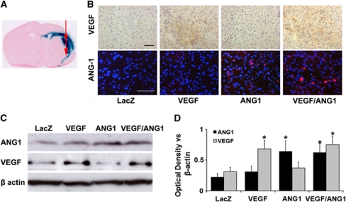
Figure 2.
Adeno-associated viral vector (AAV) mediated transgene expression. (A) A representative image of a coronal section shows LacZ expression around the injection sites at the border of the infarct area. (B) Representative images of vascular endothelial growth factor (VEGF) and angiopoietin-1 (ANG1) antibody-stained sections. VEGF expression was detected in the VEGF and VEGF/ANG1 groups (brown). ANG1 expression was detected in the ANG1 and VEGF/ANG1 groups (red). Scale bars are 100 μm. (C) Representative Western blots are shown. β-Actin was used as loading control. (D) Bar graph shows the quantification of the protein expression analyzed from Western blots. The expression of VEGF and ANG1 increased only in corresponding vector-injected brain tissues. *P<0.05 versus AAV-LacZ group, N=3 per group. LacZ: AAV-LacZ-injected group; ANG1: AAV-ANG1-injected group; VEGF: AAV-VEGF-injected group; and VEGF/ANG1: AAV-VEGF and AAV-ANG1-coinjected groups. The color reproduction of this figure is available at online version.

