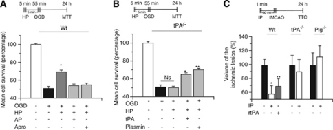
Figure 3.
Endogenous tissue-type plasminogen activator (tPA) induces hypoxic tolerance. Mean cell survival determined by the 3-(4,5-dimethylthiazol-2-yl)-2,5-diphenyltetrazolium bromide (MTT) assay in wild-type (Wt; A) and tPA-deficient (tPA−/−; B) neurons exposed to 5 minutes of oxygen–glucose deprivation (OGD) conditions (hypoxic preconditioning, HP), followed 5 minutes later by 55 minutes of OGD. A subgroup of neurons was incubated during the preconditioning phase with either α2-antiplasmin 100 nmol/L (AP) or aprotinin 1.4 μmol/L (Apro), or tPA 5 nmol/L or plasmin 10 nmol/L. HP denotes the moment when cells were exposed to sublethal hypoxia; n=12 for each observation. Data represent mean cell survival±s.d. (A) *P<0.01 versus cells exposed to OGD without hypoxic preconditioning. (B) *P<0.01 versus tPA−/− neurons preconditioned in the absence of tPA and **P<0.01 versus tPA−/− neurons preconditioned in the absence of plasmin. (C) Mean percentage decrease in the volume of the ischemic lesion compared with non-preconditioned mice (black bars) in Wt, tPA−/−, and plasminogen (Plg−/−) mice preconditioned by exposure to 1 minute of low oxygen concentration (ischemic preconditioning, IP), followed 60 minutes later by transient middle cerebral artery occlusion (tMCAO). Black bars depict mice undergoing tMCAO without preconditioning. A subgroup of Wt mice was preconditioned with recombinant tPA (rtPA) intravenously administered 60 minutes before tMCAO (gray bar); n=12 for each observation. Data represent mean percentage compared with stroke volume in non-preconditioned brains±s.d. *P<0.01 compared with non-preconditioned Wt mice. **P<0.05 versus non-preconditioned Wt mice. Ns, non significant; TTC, triphenyltetrazolium chloride staining.

