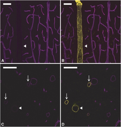
Figure 1.
P-glycoprotein (P-gp) expression is heterogeneous in cerebral vessels. P-gp/smooth muscle actin (SMA) double staining in the cortex (A, B) and hippocampus (C, D). (Left panels) (A, C) P-gp staining (magenta). (Right panels) (B, D) Merged images of P-gp (magenta) and SMA (yellow) stainings. In both the brain regions, P-gp staining is barely detectable in the largest arterioles (arrowheads) and much fainter in small arterioles (arrows) than in neighboring venules and capillaries. Bar: 30 μm.

