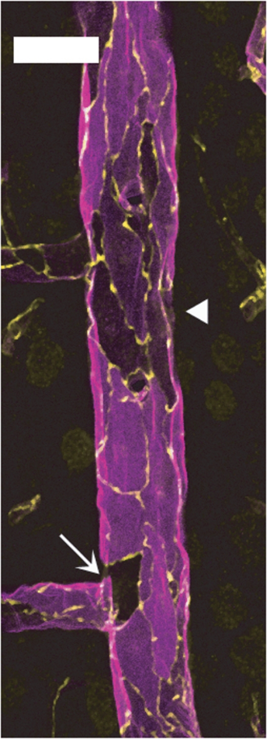
Figure 7.
Endothelial barrier antigen (EBA) expression in venular endothelium is highly heterogeneous. Longitudinal hemisection of a large cortical vein doubly labeled for EBA (magenta) and occludin (yellow). Several endothelial cells, either isolated (arrow) or clustered (arrowheads), show low or undetectable EBA staining. Bar: 30 μm

