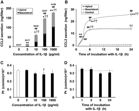
Figure 3.
Secretion of CCL2 by primary cultures of choroidal epithelial cells. The cells were seeded on Transwell Clear inserts as described in Materials and methods. The experiments were performed 5 days after the cells reached confluence (n=3 to 6 per group). The apical surface of epithelial monolayers was exposed to interleukin-1β (IL-1β). (A) Dose-response studies. The cells were exposed to IL-1β for 6 hours. The fractions above the columns represent the proportion (%) of apical versus basolateral release of CCL2 into the culture media. Under control conditions, CCL2 was constitutively produced, and the total amount of CCL2 secreted into the apical and basolateral chambers during the 6-hour observation period was 0.5 ng/filter. This secretion was polarized, with CCL2 being predominantly released across the apical (cerebrospinal fluid (CSF)-facing) membrane. Note that, with all IL-1β concentrations tested, except 1 ng/mL, the larger amounts of CCL2 were secreted into the apical chamber when compared with those released into the basolateral compartment. (B) Time-course studies. The cells were incubated with 10 pg/mL of IL-1β for up to 24 hours. Note that during the initial 6-hour period of exposure to IL-1β, the CCL2 levels in the apical and basolateral chambers increased rapidly; however, later on, the CCL2 levels either did not change (basolateral) or increased at a much slower rate (apical). (C) The permeability of epithelial monolayers to 14C-sucrose after the 6-hour incubation with varying concentrations of IL-1β. The permeability coefficient, Pt, was calculated from the 14C-sucrose clearance rates as described in Materials and methods. (D) The permeability of epithelial monolayers to 14C-sucrose after the exposure to 10 pg/mL of IL-1β for up to 24 hours. The dashed line represents the Pt value observed in untreated cells. All data represent mean values±s.d. *P<0.05, **P<0.01 for apical versus basolateral CCL2 secretion (Student's t-test with Bonferroni correction). †P<0.05, ††P<0.01 for CCL2 secretion in response to IL-1β versus control (Dunnett test). ##P<0.01 for a difference in CCL2 secretion in response to the next higher concentration of IL-1β or between two consecutive time points during the exposure to the cytokine (Tukey–Kramer test).

