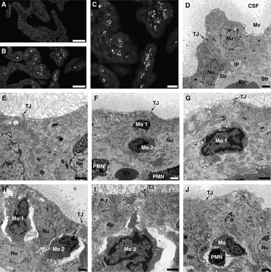
Figure 4.
Posttraumatic accumulation of monocytes in the lateral ventricle choroid plexus (CP). (A, B) Low-power microphotographs of the contralateral and ipsilateral CPs, respectively, at 1 day after traumatic brain injury (TBI). Monocytes were stained with anti-CD68 antibody. These inflammatory cells were only sporadically found in the ipsilateral CP at 6 hours post-TBI. (C) A higher magnification microphotograph of the ipsilateral CP shown in (B). (D) Transmission electron microscopic analysis of the CP. The choroidal tissue was harvested at 1 day after TBI. The contralateral CP, which has normal morphology, similar to the morphology of choroidal tissue from sham-injured rats, is shown. Note that the choroidal epithelial cells have well-defined microvilli and the lateral membrane is folded into elaborate interdigitating processes near the base of the cells. The space between adjacent epithelial cells is narrow and the tight junctions between the cells are well discernible. (E) A higher magnification microphotograph of the contralateral CP. (F) The ipsilateral CP. The microphotograph shows two monocytes (Mo 1 and Mo 2) that reached the intercellular space between the choroidal epithelial cells. Note that the space between invading monocytes and the epithelial cells is narrow. The microphotograph also shows two neutrophils or polymorphonuclear leukocytes (PMN) that extravasated into the choroidal stroma. (G) A higher magnification view of monocyte 1 from (F). (H) The ipsilateral CP. Two monocytes (Mo 1 and Mo 2) that reached the intercellular space between the epithelial cells are shown. A significant expansion of space between invading inflammatory cells and the choroidal epithelial cells is well visible on this microphotograph. Such enlarged, rather than tight, space between migrating monocytes and the epithelial cells was predominantly seen in the ipsilateral CP. (I) A higher magnification view of monocyte 2 from (H). Note that the movement of monocytes between the epithelial cells toward their apical domain does not appear to affect the integrity of tight junctions. (J) The ipsilateral CP. A monocyte and neutrophil migrating in tandem between adjacent epithelial cells. Bars: panels (A, B), 50 μm; panel (C), 20 μm; panels (D, F, H, J), 2 μm; panels (E, G, I), 1 μm. BV, blood microvessel; CSF, cerebrospinal fluid; IP, interdigitating processes; Mo, monocyte; Mv, microvilli; Nu, nucleus of the choroidal epithelial cell; PMN, neutrophil; Str, choroidal stroma; TJ, tight junction.

