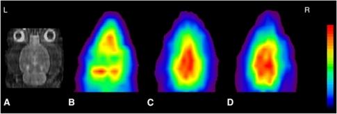
Figure 2.
Coronal magnetic resonance imaging (MRI) (A), C15O-HbV (B), H215O (C), and 15O2-HbV (D) positron emission tomography (PET) images of a normal rat. Average PET images of 2 minutes for C15O-HbV, and 3 minutes for H215O and 15O2-HbV after reaching equilibrium were used to calculate the hemodynamic parameters. Each PET image was coregistered to the individual MRI, and sliced at the same level. The upper values for the color scale were 50, 300, and 300 (kBq/mL) for (B), (C), and (D), respectively. HbV, hemoglobin-containing vesicles.

