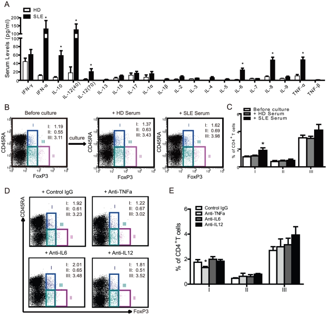
Figure 5. Proinflammatory cytokines contribute to CD45RA+FoxP3+ T cell development in SLE.
(A) A series of cytokines were measured by high sensitivity multiplex assay from the sera of healthy donors (n=15) and active SLE patients (n=15). *p<0.05 compared to healthy sera. (B) Healthy isolated PBMCs were stimulated with the sera (1∶5 dilutions) from autologous healthy donors or active SLE patients, cultured for 3 days and analyzed for CD45RA and FoxP3 expression by FCM before and after culture. Representative FCM data was shown. (C) Statistical analysis of the percentage of each FoxP3+ subset among CD4+ T cells was performed before and after culture as defined in (B) from 5 healthy donors and 5 active SLE patients. *, p<0.05 compared to unstimulated control. (D) Healthy isolated PBMCs were stimulated by the sera (1∶5 dilutions) from active SLE patients with control IgG (10 µg/ml), anti-TNFa (10 µg/ml), anti-IL6 (10 µg/ml), anti-IL12 (10 µg/ml) respectively, cultured for 3 days and analyzed for CD45RA and FoxP3 expression by FCM. Representative FCM data was shown. (E) Statistical analysis of the percentage of each FoxP3+ subset among CD4+ T cells was performed as defined in (D) from 5 active SLE patients. *, p<0.05 compared to control IgG.

