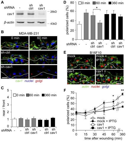
Figure 2. Polarization of metastatic cells is dependent on caveolin-1.
(A) Total protein extracts were prepared from both parental MDA-MB-231 and cells stably transduced with a control shRNA targeting luciferase (sh-ctrl) or a shRNA targeting caveolin-1 (shRNA-caveolin-1 #5, sh-cav1). Extracts were separated by SDS-PAGE (35 µg total protein per lane) and analyzed by Western Blotting with antibodies against caveolin-1 and β-actin. (B) Confluent monolayers of parental, shRNA-control (sh-ctrl) or shRNA-caveolin-1 (sh-cav1) treated MDA-MB-231 cells were wounded with a pipet tip and fixed at 0 (left panel) and 60 minutes (right panel) after monolayer injury. Samples were stained with anti-caveolin-1 (green) and anti-Golgin-97 (red) antibodies, whereas the nuclei were stained with DAPI (blue). The first layer of cells facing the wounded area is distinguished from the rest by a white line. Scale bar, 20 µm. (C) Caveolin-1 localization was analyzed as described in Figure 1C, by measuring the ratio of fluorescence intensity “rear/front”. Data are the mean ± SEM, n = 3. (D) Polarization of both parental (-) and shRNA treated MDA-MB-231 cells with shRNA-luciferase (sh-ctrl) or shRNA-caveolin-1 (sh-cav1) was measured from (C) as described in the Materials and Methods, by analyzing the outer layer of cells facing the wounded area (B). The percentage of polarized cells was evaluated at 0, 60 and 360 minutes after wounding. Data were averaged from three independent experiments (mean ± SEM). *Comparison with control shRNA P<0.05. (E) B16-F10 cells transfected with either pLacIOP (mock) or pLacIOP-caveolin-1 (cav1) were treated or not with 1mM IPTG for 24 hours, grown in monolayers and wounded with a pipet tip to allow migration for 1 hours. Samples were stained with anti-Gigantin-1 polyclonal antibody (blue), phalloidin-Alexa488® (green) and propidium iodide (red). The outer layer of cells facing the wounded area is outlined with a white line. (F) Cell polarization was measured as described in the Materials and Methods, by analyzing the outer layer of cells facing the wounded area. The percentage of polarized cells was evaluated 0-360 minutes after wounding the monolayer. Data were averaged from three independent experiments (mean ± SEM). Statistically significant differences compared with pLacIOP-transfected B16-F10 (mock) cells are indicated (**, P<0.01 at time 360 min; *, P<0.05 at time 60 min).

