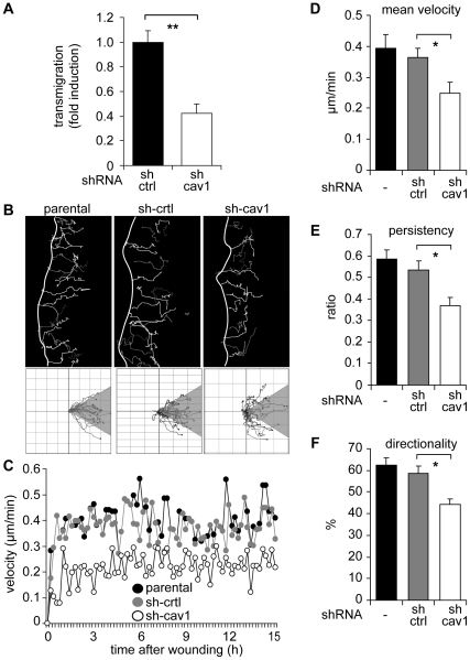
Figure 3. Caveolin-1 favors directionality and persistence of migration in MDA-MB-231 cells.
(A) Migration of MDA-MB-231 cells treated with shRNA targeting either luciferase (sh-ctrl) or caveolin-1 (sh-cav1) was assessed in a Boyden chamber assay. Cells (5×10⁴) were seeded on fibronectin-coated (2 µg/ml) transwell plates and allowed to migrate for 2 hours. Cells that migrated to the lower side were detected with crystal violet staining (mean ± SEM, n = 3). **P<0.01. (B) Parental and shRNA treated MDA-MB-231 cells were grown as confluent monolayers, wounded with a pipet tip and migration was recorded by time-lapse video microscopy (total 15 hours, 12 minutes frame interval). Cell tracks were determined by using the Image J software (“Manual Tracking” plug-in). Tracks of single cells at the wounded edge are shown (upper panel). The edge of the wound is indicated with a white line. Single cell tracks are shown in a Cartesian coordinate system for each cell type (lower panel). (C) Instant velocity was analyzed for each cell type in (B) and plotted as a function of time (0-15 hours). (D) Mean velocity values were obtained from (C) and plotted for each cell type. (E) Persistency of migration was calculated as the ratio between the net distance and the total distance of migration in (B). Average values were obtained for parental, sh-ctrl and sh-cav1 cells (mean ± SEM). (F) Directionality of migration was obtained from the analysis shown in (B), lower panels. Tracks that lay within a 60° angle with respect to the direction of cell movement were considered as oriented (shaded region). The percentage of cells with orientated tracks was calculated and plotted (mean ± SEM, n = 3). *P<0.05.

